Which Ventricles Are Divided by the Septum Pellucidum
Third and fourth ventricles. Where does the 4th ventricle start.
Ventricles Of The Brain Labeled Anatomy Function Csf Flow Definition Ezmed
Septum Pellucidum in Coronal Projection.
. It is signifi-cantly more common in women3. The septum pellucidum is a small membrane wall in the brain of humans and some animals. The lateral ventricles are cavities within the cerebrum.
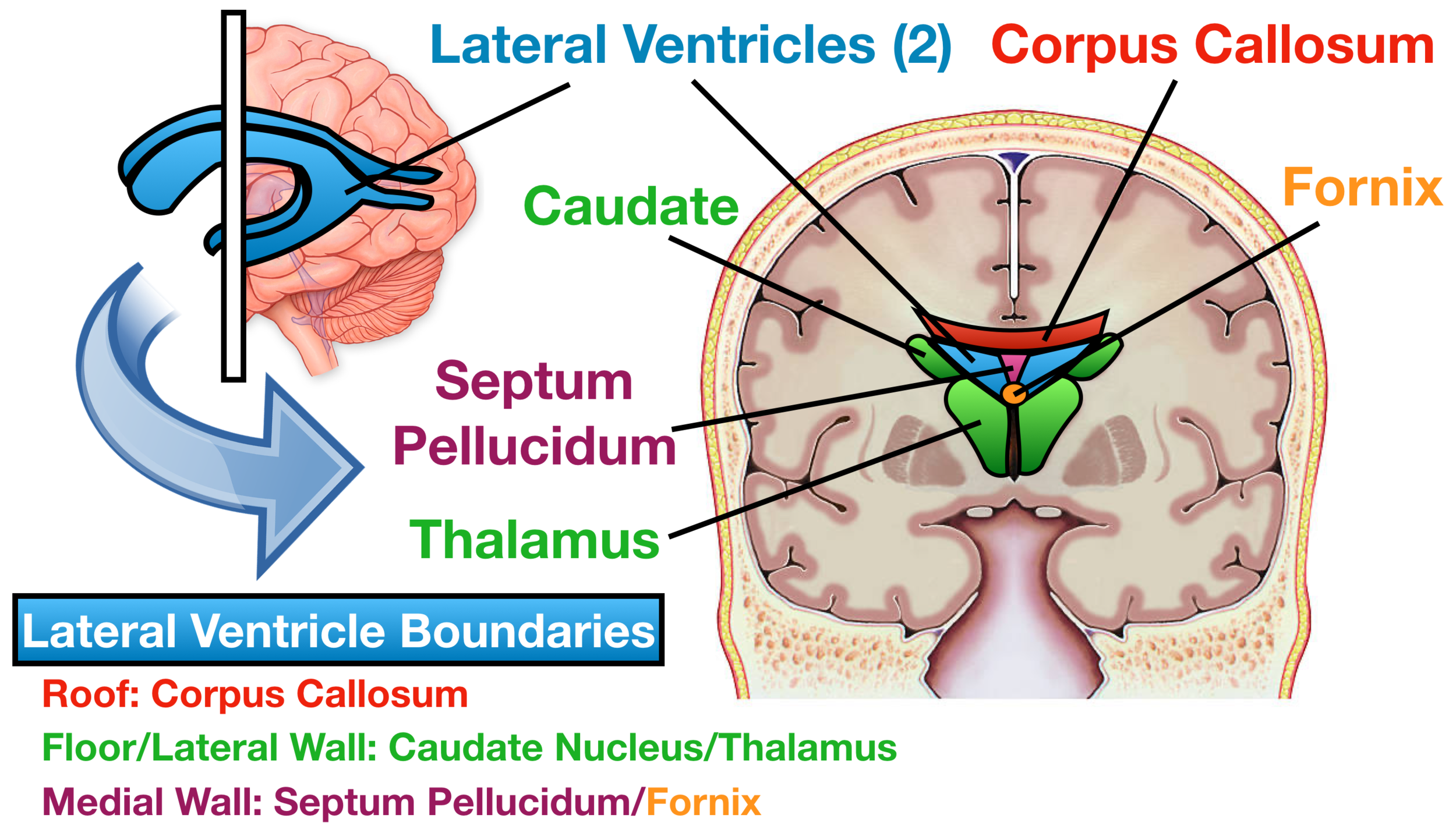
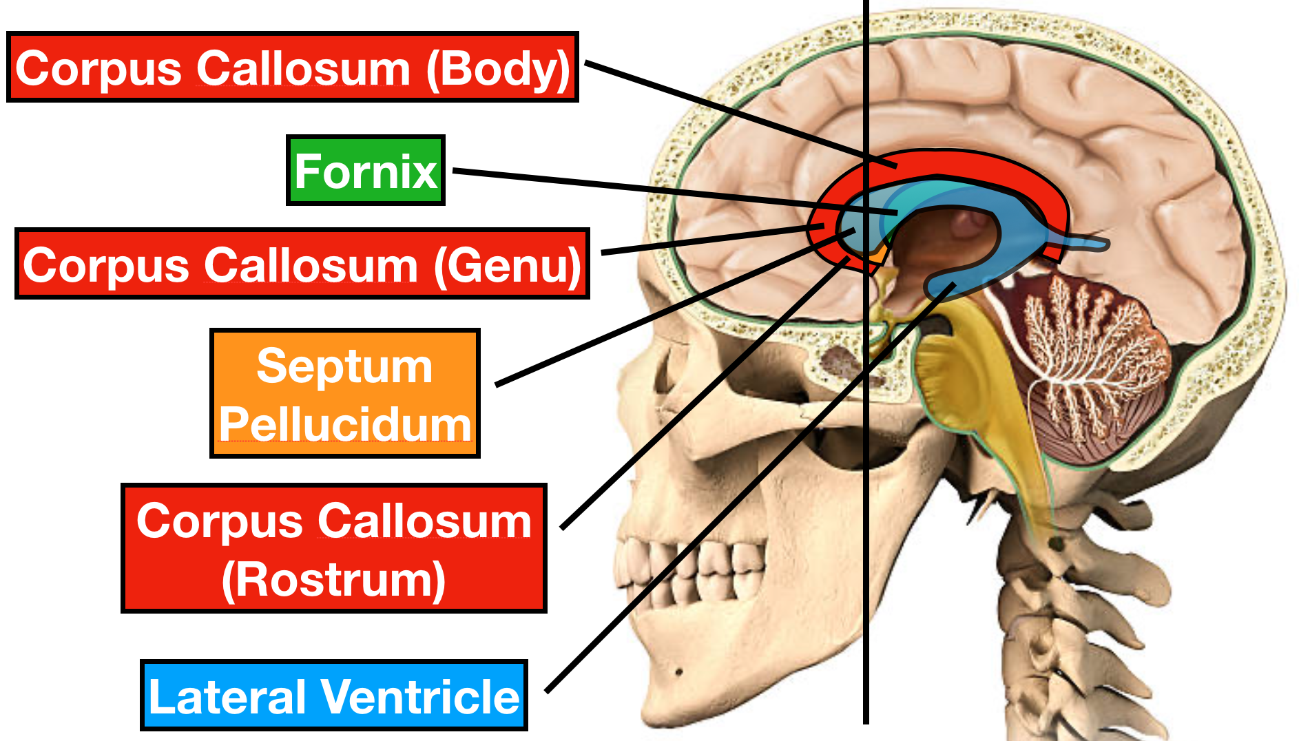
The septum pellucidum is lined with ependymal cells on its ventricular side whilst pia mater lines the cavity. In terms of location it extends from the corpus collosum a collection of neural fibers under the cortex. The septum pellucidum is a transparent membrane that divides the right and left lateral ventricles in the middle of the body.
The cavum septum pellucidum CSP is a potential cavity between the membranous leaves of the septum pellucidum separated by at least 1 mm and is considered a normal anatomical variation. They are easily recognized as a T shaped midline structure divided in the middle by the gracile septum pellucidum. 1 Cavum septum pellucidum.
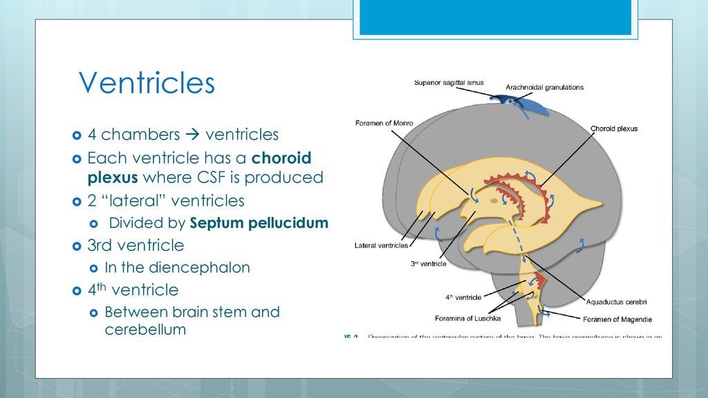
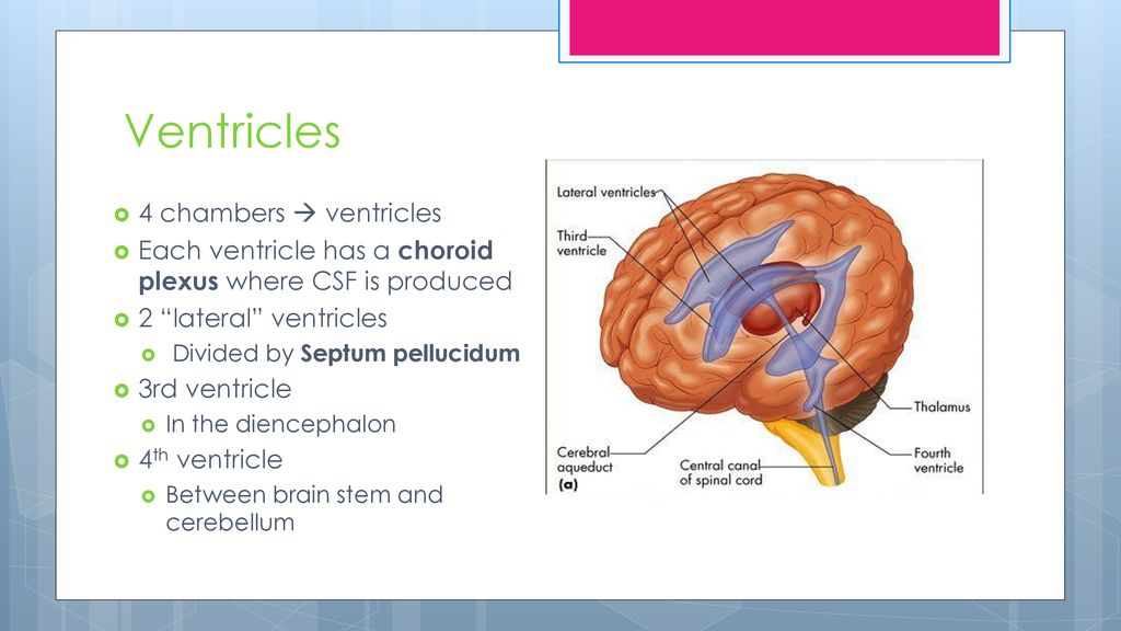
The ventricles are all interconnected. What type of tissue makes up the cerebral cortex. The frontal horns are seen in this coronal MRI using a STIR sequence.
The septum pellucidum Latin for translucent wall is a thin triangular vertical double membrane separating the anterior horns of the left and right lateral ventricles of the brain. Anterior horns and body of lateral ventricles. White matter has a fatty consistency.
It runs as a sheet from the corpus callosum down to the fornix. The composition of gray matter includes neuron cell bodies. Septo-optic dysplasia This disorder occurs in early brain development and also includes underdevelopment of.
Which ventricles are divided by the septum pellucidum. The right and left lateral ventricles are. Lateral and third ventricle.
It is made up of a thin two-layered structure that consists of white. Which ventricles are divided by septum pellucidum. Its main job is to separate the lateral ventricles important passageways within the brain tissue and the membrane forms whats basically a barrier between these sections.
The frontal horns course through the frontal lobes and above the anterior portion of the temporal lobes. -surround the capillaries of the choroid plexus. It extends between the anterior portion of the corpus callosum and the body of the fornix and its width varies from 15 to.
The septum pellucidum meaning translucent wall in Latin - SP also known as the ventricle of Sylvius is a thin triangular double membrane separating the frontal horns of the right and left lateral ventricles of the brain. Cerebrospinal fluid is produced within the ventricles. What is the function of white matter.
-remove waste products from CSF. First and second ventricles. It is also known as cavum septi pellucidi the ventricle of the septum or fifth ventricle fig.
What ventricles are divided by the septum pellucidum. The septum pellucidum is a skinny partition in the center of the two lateral ventricles that separates them from one another. Fourth ventricle Fornix Lateral ventricles Third ventricle.
Hydrocephalus or water on the brain may result from blockage of CSF circulation or excessive CSF production. Which of the following ventricles is found under the corpus callosum. The thin partition that separates the first and second ventricles is the -septum insula.
Which ventricles are divided by the septum pellucidum. It also contains scattered glial cells nerve fibers and veins 1-2. Identify the passageway found in the spinal cord that is continuous with the ventricles.
The septum pellucidum acts as a partition between a portion of the lateral ventricles forming part of the walls of the anterior region of the lateral ventricles. It can be found in all fetuses at 36 weeks ges-tation and persists in 36 of full-term infants only 6 of them persist after the six month of live3411-13. SVs of both sides were evaluated according to number size distribution and location relative to.
CSP and CV were respectively and incorrectly called the fifth and sixth ventricles in the past. As a result of there are two lateral ventricles the ventricle in the diencephalon is known as the third ventricle. What type of tissue makes up the cerebral cortex.
Which ventricles are divided by the septum pellucidum. The septum pellucidum was exposed bilaterally and divided into 3 regions. Secrete CSF into the ventricles adjust the composition of CSF surround the capillaries of the chroid plexus remove waste products from CSF statement regarding cerebrospinal fluid if CSF is not properly reabsorbed the result would be hydrocephalus.
-secrete CSF into the ventricles. The lateral ventricles are chambers within the cerebrum that contain many organs.
Anatomy Of The Brain Ppt Download
Ventricles Of The Brain Labeled Anatomy Function Csf Flow Definition Ezmed
Brain Cranial Nerves Ppt Download
No comments for "Which Ventricles Are Divided by the Septum Pellucidum"
Post a Comment